42 diagram of the eye with labels
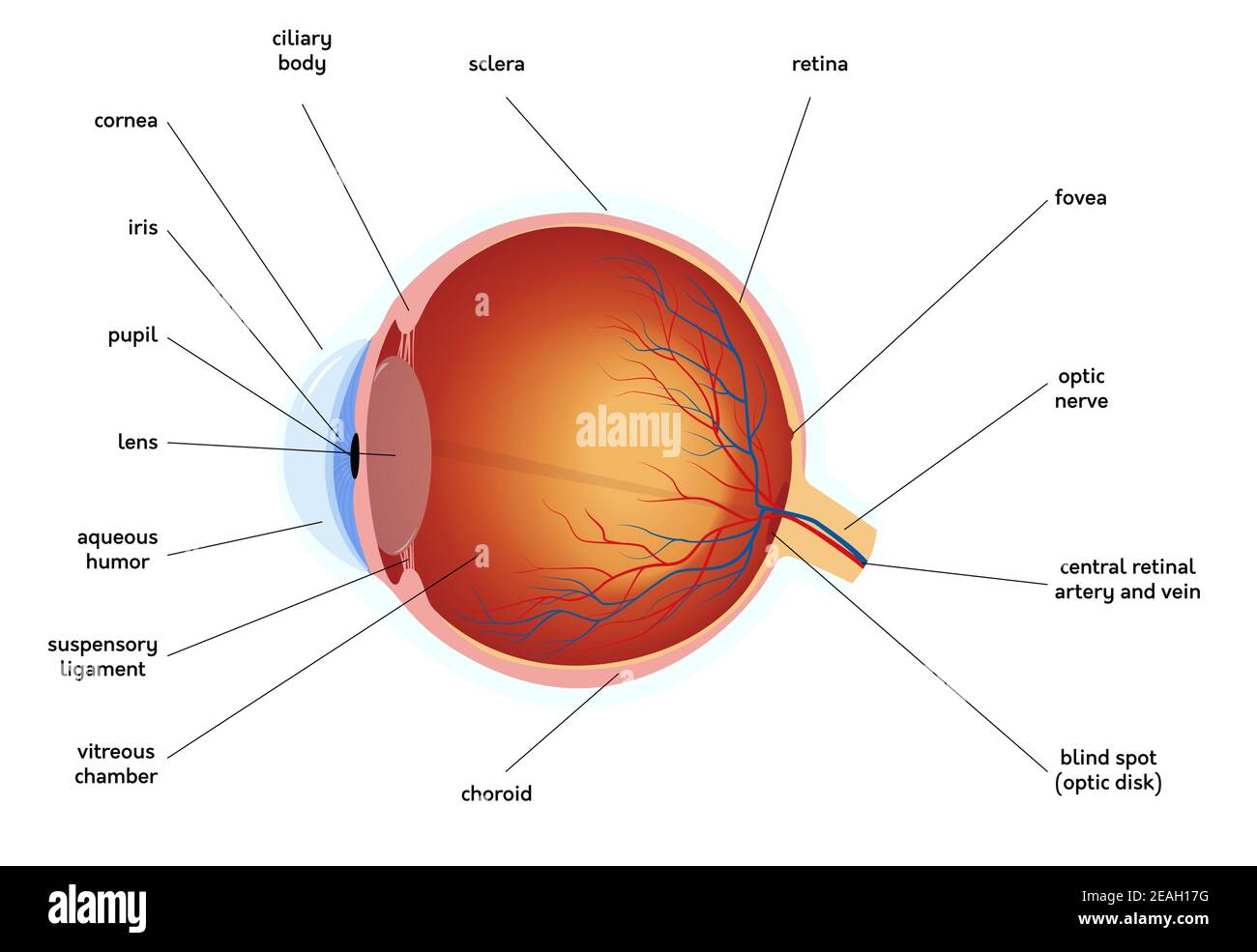
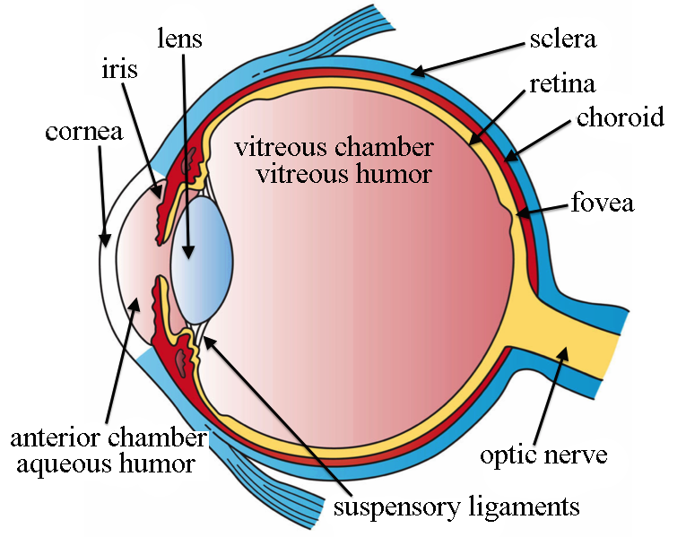
File : Schematic diagram of the human eye en.svg - Wikimedia Diagram of the human eye in English. It shows the lower part of the right eye after a central and horizontal section. Date: 25 January 2007: ... Full redraw: Group labels in accordance with the "Foundational Model Explorer." Added "Macula" and "Uvea" and removed "Zonular fibres." It has given better visibility inside the eye. Diagram of the Eye - Lions Eye Institute The eye - one of the most complex organisms in the human body. It is made up of many different parts working in unison together. In order for the eye to work at its best, all parts must work well collectively. To understand the eye and its functions, it's important to understand how the eye works, see below diagrams for both the external ...
Label the Eye Worksheet - Teacher-Made Learning Resources - Twinkl In this resource, you'll find a 2-page PDF that is easy to download, print out, and use immediately with your class. The first page is a labelling exercise with two diagrams of the human eye. One is a view from the outside, and the other is a more detailed cross-section. Challenge learners to label the parts of the eye diagram. On the second page, you'll find a set of answers showing the ...
:max_bytes(150000):strip_icc()/GettyImages-695204442-b9320f82932c49bcac765167b95f4af6.jpg)
Diagram of the eye with labels
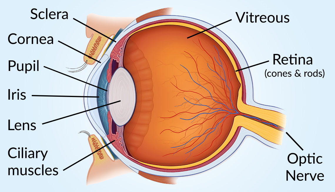
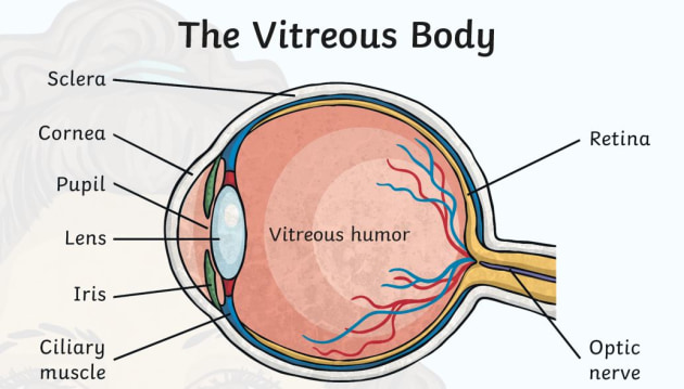
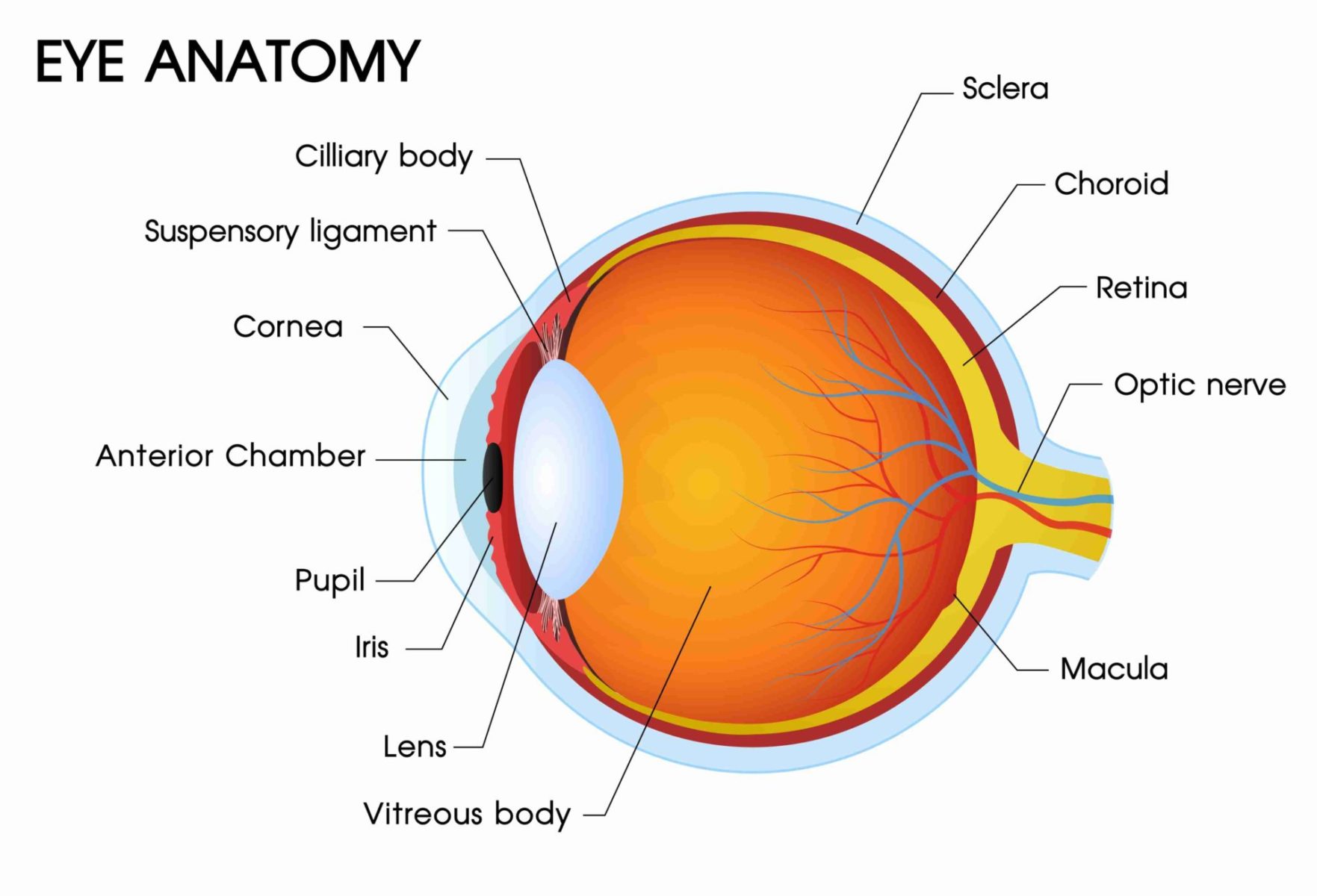
Labeled Eye Diagram - Science Trends The human eye is composed of many different parts that work together to interpret the world around us. What you want to interpret as a major part of the human eye is somewhat up to the individual, but in general there are seven parts of the human eye: the cornea, the pupil, the iris, the lens, the vitreous humor, the retina, and the sclera. Let's take a closer look at each of these ... Anatomy of the eye: Quizzes and diagrams | Kenhub Take a look at the diagram of the eyeball above. Here you can see all of the main structures in this area. Spend some time reviewing the name and location of each one, then try to label the eye yourself - without peeking! - using the eye diagram (blank) below. Unlabeled diagram of the eye. Click below to download our free unlabeled diagram of ... Structure and Functions of Human Eye with labelled Diagram - BYJUS The External Structure of an Eye. Sclera: It is a white visible portion. It is made up of dense connective tissue and protects the inner parts. Conjunctiva: It lines the sclera and is made up of stratified squamous epithelium. It keeps our eyes moist and clear and provides lubrication by secreting mucus and tears.
Diagram of the eye with labels. Anatomy of the Eye | Johns Hopkins Medicine The back part of the eye's interior. Pupil. The opening in the middle of the iris through which light passes to the back of the eye. Retina. The light-sensitive nerve layer that lines the inside of the back of the eye. The retina senses light and creates impulses that are sent through the optic nerve to the brain. Sclera. Eye Anatomy: Parts of the Eye and How We See Here is a tour of the eye starting from the outside, going in through the front and working to the back. Eye Anatomy: Parts of the Eye Outside the Eyeball. The eye sits in a protective bony socket called the orbit. Six extraocular muscles in the orbit are attached to the eye. These muscles move the eye up and down, side to side, and rotate the eye. Labelling the eye — Science Learning Hub Labelling the eye. Use this interactive to label different parts of the human eye. Drag and drop the text labels onto the boxes next to the diagram. Selecting or hovering over a box will highlight each area in the diagram. The human eye has several structures that enable entering light energy to be converted to electrochemical energy. eye labeling Diagram | Quizlet It is the first structure to refract (bend) light that enters the eye. sclera. Tough white out covering of the eyeball. choroid. Middle layer of the eye (between the retina and the sclera) that contains the blood vessels that nourish the eye and cornea ... Label the Ear. 12 terms. aimige. Recent flashcard sets. ACC 4400 Practice Ch 12. 36 terms ...
Labelled Diagram of Human Eye, Explanation and Function - VEDANTU Labeled Diagram of Human Eye . The eyes of all mammals consist of a non-image-forming photosensitive ganglion within the retina which receives light, adjusts the dimensions of the pupil, regulates the availability of melatonin hormones, and also entertains the body clock. The Eyes (Human Anatomy): Diagram, Optic Nerve, Iris, Cornea ... - WebMD The front part (what you see in the mirror) includes: Iris: the colored part. Cornea: a clear dome over the iris. Pupil: the black circular opening in the iris that lets light in. Sclera: the ... File : Diagram of human eye without labels.svg - Wikimedia File:Diagram of human eye without labels.svg. File. : Diagram of human eye without labels.svg. Size of this PNG preview of this SVG file: 410 × 430 pixels. Other resolutions: 229 × 240 pixels | 458 × 480 pixels | 732 × 768 pixels | 976 × 1,024 pixels | 1,953 × 2,048 pixels. Eye Anatomy: A Closer Look At the Parts of the Eye - All About Vision Eye anatomy: A closer look at the parts of the eye. When surveyed about the five senses — sight, hearing, taste, smell and touch — people consistently report that their eyesight is the mode of perception they value (and fear losing) most. Despite this, many people don't have a good understanding of the anatomy of the eye, how vision works ...
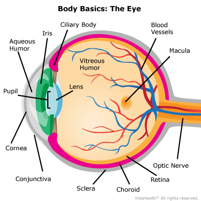
What Does the Eye Look Like? - Diagram of the Eye | Harvard Eye Associates It is mostly water and gives the eye its form and shape. Our eyes are vital for seeing the world around us. Keep them healthy by maintaining regular vision exams. Contact Harvard Eye Associates at 949-951-2020 or harvardeye.com to schedule an appointment today. Eye Diagram With Labels and detailed description - BYJUS The Cornea is a dome-shaped tissue covering the front of the eye. Iris is the coloured part of the eye and controls the amount of light entering the eye by regulating the size of the pupil. The lens is located just behind the iris. Its function is to focus the light on the retina. The optic nerve transmits electrical signals from the retina to ... Eye Anatomy: 16 Parts of the Eye & Their Functions - Vision Center The following are parts of the human eyes and their functions: 1. Conjunctiva. The conjunctiva is the membrane covering the sclera (white portion of your eye). The conjunctiva also covers the interior of your eyelids. Conjunctivitis, often known as pink eye, occurs when this thin membrane becomes inflamed or swollen. Label Parts of the Human Eye - University of Dayton Parts of the Eye. Select the correct label for each part of the eye. The image is taken from above the left eye. Click on the Score button to see how you did. Incorrect answers will be marked in red. ...
Structure and Function of the Human Eye
Human Eye Ball Anatomy & Physiology Diagram - eMedicineHealth The orbit is the bony eye socket of the skull. The orbit is formed by the cheekbone, the forehead, the temple, and the side of the nose. The eye is cushioned within the orbit by pads of fat. In addition to the eyeball itself, the orbit contains the muscles that move the eye, blood vessels, and nerves. The orbit also contains the lacrimal gland ...

New way to draw a human eye and label its parts
PDF Parts of the Eye - National Institutes of Health Eye Diagram Handout Author: National Eye Health Education Program of the National Eye Institute, National Institutes of Health Subject: Handout illustrating parts of the eye Keywords: parts of the eye, eye diagram, vitreous gel, iris, cornea, pupil, lens, optic nerve, macula, retina Created Date: 12/16/2011 12:39:09 PM

Human Eye Diagram | Label Diagram Of Eye | Science Drawing
Structure and Functions of Human Eye with labelled Diagram - BYJUS The External Structure of an Eye. Sclera: It is a white visible portion. It is made up of dense connective tissue and protects the inner parts. Conjunctiva: It lines the sclera and is made up of stratified squamous epithelium. It keeps our eyes moist and clear and provides lubrication by secreting mucus and tears.
Eye diagram labels - Teaching resources
Anatomy of the eye: Quizzes and diagrams | Kenhub Take a look at the diagram of the eyeball above. Here you can see all of the main structures in this area. Spend some time reviewing the name and location of each one, then try to label the eye yourself - without peeking! - using the eye diagram (blank) below. Unlabeled diagram of the eye. Click below to download our free unlabeled diagram of ...

Module 1: Labeled Diagram of the Eye | Diagram of the eye ...
Labeled Eye Diagram - Science Trends The human eye is composed of many different parts that work together to interpret the world around us. What you want to interpret as a major part of the human eye is somewhat up to the individual, but in general there are seven parts of the human eye: the cornea, the pupil, the iris, the lens, the vitreous humor, the retina, and the sclera. Let's take a closer look at each of these ...

Draw a diagram of vertical section of human eye and label the ...

Eye labeling Diagram | Quizlet

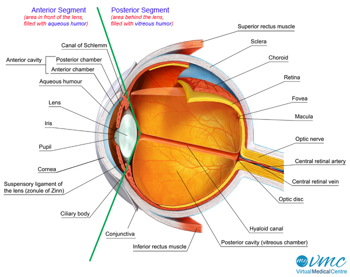
Label the eye parts correctly.i Aqueous humour ii vitreous ...
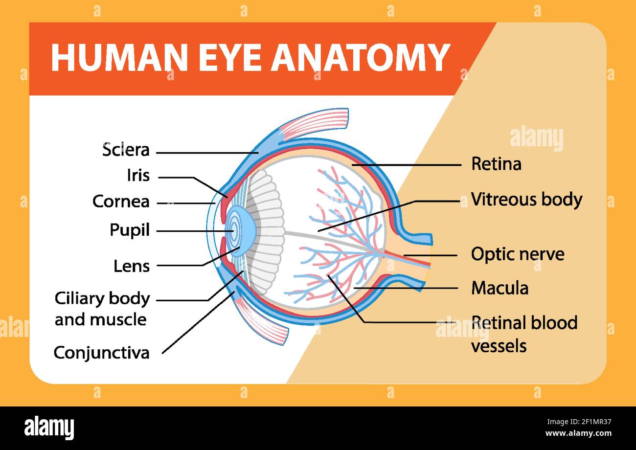
Diagram of human eye anatomy with label illustration Stock ...

Sensory Structures | BioNinja

13,693 Eye Diagram Images, Stock Photos & Vectors | Shutterstock

How the eye works | RNIB
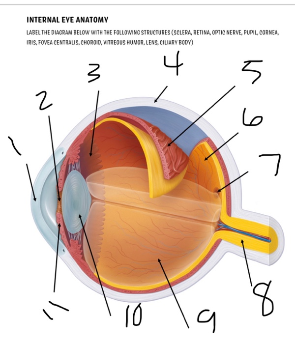
Solved INTERNAL EYE ANATOMY LABEL THE DIAGRAM BELOW WITH THE ...

3d Image Render Of Diagram Of Eye Anatomy With Label For ...

Chapter 9a: Internal Lateral view of the Eye Diagram | Quizlet

Vector Illustration Of Diagram Of Eye Anatomy With Label ...

Anatomy of the Human Eye
Vision and Eye Diagram: How We See

File:Schematic diagram of the human eye en.svg - Wikimedia ...

Simple eye diagrams | Easy eye diagram | Labeled eye diagram ...

Diagram Of Eye For Kids The Eye Diagram For Kids Nurse ...

Sensory Structures | BioNinja

Draw a labelled diagram of human eye and explain the image of ...

Draw a labelled diagram of the human eye Label the following ...
Vision and the eye's anatomy | HealthEngine Blog
Labelling the eye - Teaching resources

Label Parts of the Human Eye

eye labeling Diagram | Quizlet
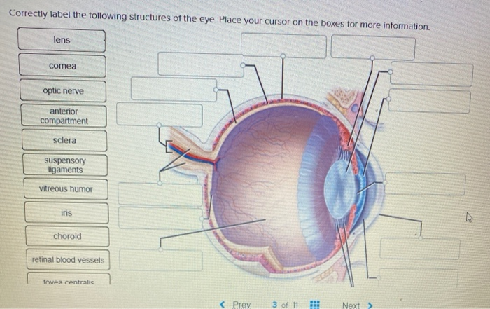
Solved Correctly label the following structures of the eye ...
Eye diagram labels - Teaching resources

Anatomy of the Eye | Red Rover Ventures
Eye diagram labels - Teaching resources
Pupil eye diagram hi-res stock photography and images - Alamy
Human Eye Diagram For Kids – Fun Human Body Facts - Twinkl

Diagram of human eye anatomy with label illustration Stock ...

Diagram of human eye anatomy with label illustration. | CanStock

GCSE Biology eye labelling Diagram | Quizlet
Your Eyes (for Kids) - Nemours KidsHealth
What Does the Eye Look Like? – Diagram of the Eye | Harvard ...
Eye Anatomy: The 9 Main Parts of the Eye | Specialty Eye ...
Eye Anatomy: Parts of the Eye and How We See - American ...

Diagram of human eye anatomy with label 1945551 Vector Art at ...
:max_bytes(150000):strip_icc()/GettyImages-695204442-b9320f82932c49bcac765167b95f4af6.jpg&description=42 diagram of the eye with labels)
Post a Comment for "42 diagram of the eye with labels"